

Please use this identifier to cite or link to this item:
http://www.alice.cnptia.embrapa.br/alice/handle/doc/1041808| Title: | Scanning electron microscopy of superficial white onychomycosis. |
| Authors: | ALMEIDA JUNIOR, H. L. de  BOABAID, R. O. TIMM, V. SILVA, R. M. e. CASTRO, L. A. S. de |
| Affiliation: | Hiram Larangeira de Almeida Jr, UFPEL; Roberta Oliveira Boabaid, UCPEL; Vitor Timm, UFPEL; Ricardo Marques e Silva, UFPEL; LUIS ANTONIO SUITA DE CASTRO, CPACT. |
| Date Issued: | 2015 |
| Citation: | Anais Brasileiros de Dermatologia, v. 90, n. 5, p. 753-755, 2015. |
| Description: | Superficial white onychomycosis is characterized by opaque, friable, whitish superficial spots on the nail plate. We examined an affected halux nail of a 20-year-old male patient with scanning electron microscopy. The mycological examination isolated Trichophyton mentagrophytes. Abundant hyphae with the formation of arthrospores were found on the nail?s surface, forming small fungal colonies. These findings showed the great capacity for dissemination of this form of onychomycosis. |
| Thesagro: | Micose |
| NAL Thesaurus: | scanning electron microscopy |
| Keywords: | Unha |
| DOI: | DOI: http://dx.doi.org/10.1590/abd1806-4841.20154136 |
| Type of Material: | Artigo de periódico |
| Access: | openAccess |
| Appears in Collections: | Artigo em periódico indexado (CPACT) |
Files in This Item:
| File | Description | Size | Format | |
|---|---|---|---|---|
| LuisSuitawhiteonychomycosis.pdf | 320,04 kB | Adobe PDF | 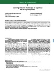 View/Open |


